I think Tuesdays are good days for cross sections. Don’t you feel that urge to perform thin slicing of plant tissue on Tuesdays? Seriously, what could be better than identifying monocots and dicots through vascular bundle arrangements!? I can’t think of any other way I’d rather spend my Tuesday. So lucky for me, this week in Oregon State plant structures lab we did exactly that! By looking at the location of the vascular bundles in plant stems, we were able to identify the plant species as monocot or dicot, assuming this wasn’t already known. Taking a close look at the vascular bundles, distinguishing of the individual components of the vascular bundles as well as the surrounding tissue was pretty clear with proper stain types and methods. Corn (Zea mays) is monocot and is in figure 1 and 2 below. TBO stain was used to provide contrast between cell types depending on the compounds present in the cell. Staining with TBO or Toluidine Blue O, will stain pectin substances pink to reddish purple, and lignin, blue or blue green. The vascular bundles are clearly stained blue which is due to the lignin of the secondary cell wall of the tracheid’s and vessel elements, the components of xylem. Phloem is the other transport tissue type found in the vascular bundles, which is composed of sieve tube elements and its dependable loving friend, the companion cells. I remember xylem and phloem and the way materials generally flow through them by saying 'xylem', in a high, squeaky voice, making me think up, and 'phloem', in a low deep voice for down. Say these words out loud a few times and hopefully it sticks in your memory for years to come like it has for myself. The xylem, transports water and minerals from the roots to the shoots and phloem carries sugars, nutrients, lipids, organics, and sad but true, viruses on a bad day. Surrounding the xylem and phloem is a sheath of sclerenchyma which helps with support and also stains thanks to its lignin found it in. Figure 1 and 2 below both help in identifying the regions referenced above.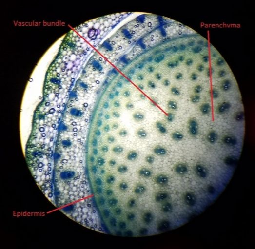 Figure 1 is a corn stem (Zea mays) cross section with TBO staining. In the image you can see the vascular bundles spread throughout the stem with parenchyma cells filling the space between the bundles and the epidermis encasing the both of them. Because the stem cross section was taken near a shoot, the tissue surrounding the epidermis of the stem is young leaf tissue. This image was taken at 40x magnification with a compound microscope. (Prepared by Lucas and photographed by Taylor) 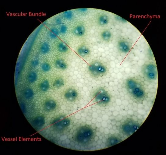 Figure 2 is a corn stem (Zea mays) cross section with TBO staining. This image is actually a more zoomed view of the same image above in figure 1. The larger cells in the vascular bundles are vessel elements. You can see two on the outside of the bundle and one or so at the bottom or top depending on orientation. The one or so at the top or bottom with the darker stained outer walls are dead and have possibly been filled with air making them look like bubbles in the slide. Vessel elements are larger transport tubes than the tracheid’s found in the xylem and are prone to air bubbles if breaks in the water tension occurs. This image was taken at 100x magnification with a compound microscope. (Prepared by Lucas and photographed by Taylor) The meristematic regions of the plant are where the new tissue is formed. There are many locations in the plant this is essentially happening. In the figures 1 and 2 above, a region running through the vascular bundles termed, procambium, creates new cells through mitosis. The procambium promotes radial growth of the plant by providing new vascular tissue to replace the non-functioning xylem and phloem. The secondary xylem and phloem get pushed away from the vascular cambium as primary vascular tissue is created giving the stem girth over time. While the pro-cambium provides lateral growth the apical meristem found at the growing tips of plants, (roots and shoots) generates upward and downward growth. Below in figure 3, you can see where the growth is taking place and the name of the region this is occurring. 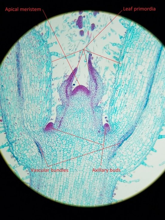 Figure 3 is a prepared slide of longitudinal section of a Coleus shoot meristem. Staining was used, which is apparent in the dividing cells (purple). In the image the leaf primordia, apical meristem, axillary buds, and vascular bundle are all visible. The image was taken at 40x on a compound microscope. (Pre prepared and photographed by Taylor) 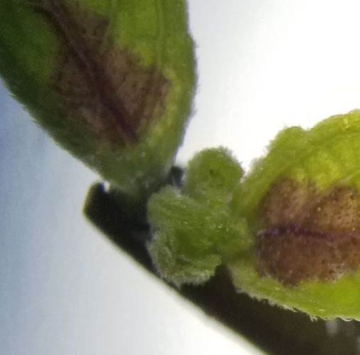 Figure 4 is an image under a dissecting microscope of a Coleus shoot meristem. In the shoot apical meristem cells are dividing by mitosis and forming new daughter cells that have yet to undergo differentiation. The swelling of these primary cell vacuoles will cause the shoot to move upwards causing primary growth. At some point these cells will differentiate into dermal, vascular, or ground tissue systems. (Prepared by John and photographed by Taylor) - Taylor Bates
0 Comments
|
AuthorContent is created by students participating in the Plant Structure course at Oregon State University for Winter 2017. Archives
March 2017
Categories
All
|
 RSS Feed
