Submitted by Brandon Quann
Hello and welcome to my very first blog post ever. I’ve decided to add my own twist to my post so I've included some of my favorite Kanye West instrumentals to add to the "ambiance" of my post, to start the playlist press play down below.
This week in lab we learned about the inner workings of seeds, seedlings, and plant cells. We carried out experiments by conducting dissections, staining plant tissues with various biological stains, preparing and viewing microscope slides, and illustrating our findings. In our lab we observed numerous plants and tissues types, however I will be focusing on our experiments with Elodea cells as well as plastids within bell peppers.
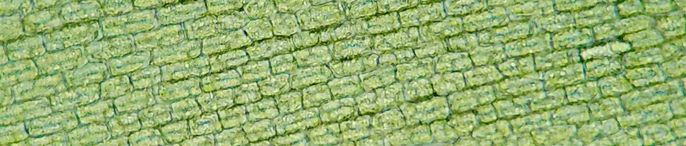
Elodea cells under 100x magnification. Photo by Taylor Bates
Who knew pondweed could be so interesting?
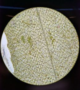
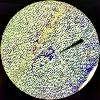
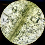
It has been quite some time since my high school biology class so when I saw that we would be studying Elodea cells in lab this week I had a trip down memory lane to my first encounter with the plant. Our observations began with making a simple wet mount of the leaf and viewing it under 100x and 400x magnification. Once we had our slides made we began searching for and sketching trichomes, chloroplasts, nuclei, vacuoles, cell walls, and any other organelles we could find.
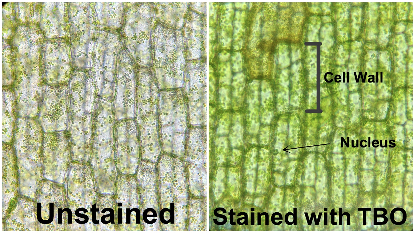
Elodea leaves were then stained with a different biological stain called Toluidine Blue O (TBO). TBO creates a variety of colors when in contact with different tissue types, the structures/ organelles and their corresponding stain color are in the following list: pectin found in cell walls, cell wall tissue, and nuclei. Based on these corresponding colors it is apparent that most abundant tissue found in Elodea leaves is from the cell wall (see photo above). A few nuclei are also visible from the stain however this is the first time I have used TBO so I am not sure if I allowed the stain enough time to work to its full potential.
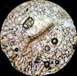
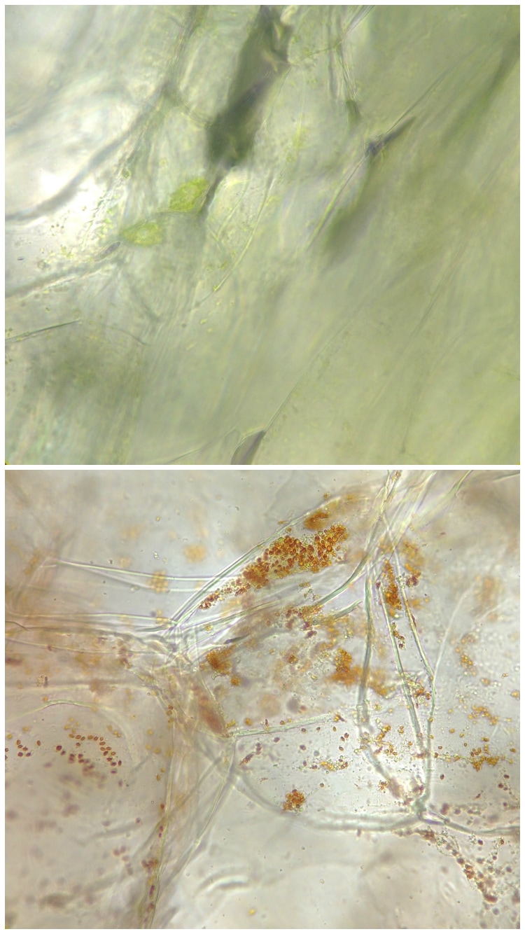
How much do Plastids cost?
dependent on environmental conditions or maturity of the fruit. These 3 different types of plastids have their own individual phenotypic differences as well as different functional roles in the cell. The photo above on the left shows the tissue of a green bell pepper with high levels of chloroplasts, these chloroplasts are the main site of photosynthesis and appear green because of the chlorophyl inside. However if the fruit is left on the plant to mature for a longer period of time the chloroplasts will be converted into chromoplasts. These chromoplasts are non-photosynthetic and work to synthesize and store pigments called carotenes, which are yellow, orange, or red in color (depending on how long they are left to mature). The conversion of chloroplasts into chromoplasts can be seen in the photo above on the right which shows a tissue sample of a red bell pepper full of red chromoplasts.
0 Comments
|
AuthorContent is created by students participating in the Plant Structure course at Oregon State University for Winter 2017. Archives
March 2017
Categories
All
|
 RSS Feed
