|
This week we looked at a few leaves belonging to plants that have adapted to survive the niche in which they found themselves. Waterlilies have adapted to survive floating atop bodies of water, the rubber plant has adapted to survive tropical and equatorial climates, and the oleander has developed advantages for surviving in very dry climates.
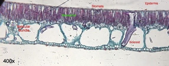
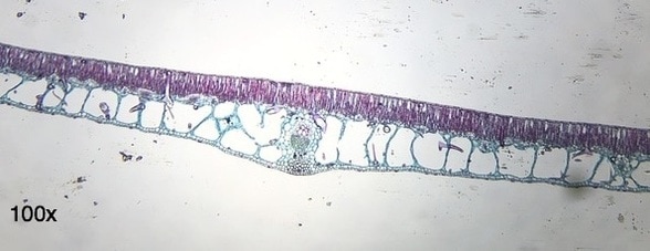
The Nymphaea (waterlily) leaf has stomata on top of their leaves instead of below to allow for increased air exchange and nutrient exchange; they do not have a defense against transpiration as other plants do in the form of guard cells. The loss due to transpiration is not a primary issue for the waterlily since it has extremely good access to water. The sclereid labeled above is meant as a support for the leaf; it helps tent up the leaf which allows for greater air exchange in the leaf air space and provides flotation for the pad. The cuticle of the leaf is quite thin and helps repel water from the stomata. -Chris Barrett 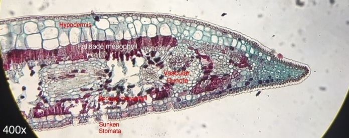 Ficus leaf, transverse section, Hematoxylin stain, 400x Magnification. Prepared by unknown and photographed by Chris Barrett 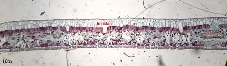 Ficus leaf, transverse section, Hematoxylin stain, 100x Magnification. Prepared by unknown and photographed by Chris Barrett The sunken stomata of the Ficus plant helps the plant retain water. By not being flush with the rest of the epidermis, the stomata allow water vapor to be released yet not be immediately blown away by wind, therefore retaining an amount of water for reabsorption. The hypodermis is quite thick on top of the palisade mesophyll cells, perhaps for protection from intense UV radiation. The sub-stomatal regions are quite large, allowing for greater gas exchange in the mesophyll cells. There are not any trichomes visible on this leaf section. This large open areas in the spongy mesophyll, the sunken stomata, and the thickened hypodermis point to this plant being able to survive very hot, and very sunny climates, perhaps in the equatorial region of the world. -Chris Barrett 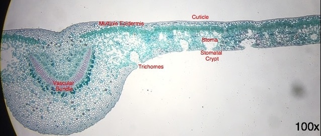 Nerium oleander leaf, transverse section, TBO stain, 100x Magnification. Prepared by unknown and photographed by Chris Barrett The adaptations of the Nerium oleander plant have allowed it to survive in very dry climates. These are evident through the presence of stomatal crypts, which contain multiple stoma positioned far away from outer line of the lower epidermis. There are also trichomes present near the openings of the stomatal crypts. Both the presence of the stomatal crypts and the trichomes located inside of them point to adaptations for survival in very dry climates. The increased presence of plant fibers in the leaves allows the leaf to maintain its shape even when its other cells are plasmolyzed in dry spouts. Its multiple epidermis and thick cuticle allows the plant to handle high ultra violet radiation.
Regardless of the fact that every part of the plant is toxic to humans and other animals, it is one of the most widely grown plants in the world due to its drought-resistance with uses including ornamental, medicine, and wind-blocking. The oleander plant is also the official flower of the city of Hiroshima as it was the first plant to flower there after the destruction of the city by nuclear blast. -Chris Barrett
0 Comments
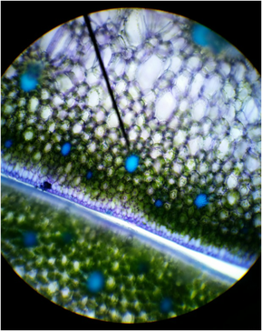 Figure 1: Cross-section of snake plant (Sansevieria trifasciata) stained with TBO, Magnification :100x, Shows sclerenchyma fibers, thickened secondary wall, parenchyma cells, and epidermis cells; Photo credit: Chris (Armstrong Plant Biology Lab) Slide prep: Chris (Armstrong Plant Biology Lab) Figure 1: Cross-section of snake plant (Sansevieria trifasciata) stained with TBO, Magnification :100x, Shows sclerenchyma fibers, thickened secondary wall, parenchyma cells, and epidermis cells; Photo credit: Chris (Armstrong Plant Biology Lab) Slide prep: Chris (Armstrong Plant Biology Lab) Introduction This past week in lab was all about learning and exploring the simple and complex tissues of plants. Our objectives were to recognize the three tissue systems of the plant body (ground, vascular, and dermal tissues), compare and contrast parenchyma, collenchyma, and sclerenchyma, identify water-conducting cells of the vascular tissue system and relate their structural features with their functions, and describe the characteristics of the epidermis, which we consider as a complex tissue. My primary focus for this lab was to prepare slides and observe the sclerenchyma fibers of a snake plant (Sansevieria trifasciata) by taking a cross-section and logitudinal-section of a leaf and observe the brachysclereids and tracheary elements of a wax pant (Hoya carnosa). Sclerenchyma Fibers of Snake Plant (Sansevieria trifasciata), Cross Section In order to examine the sclerenchyma fibers, a leaf was taken off of the snake plant. Using a razor blade, several thin cross-sections were taken from the leaf. A cross-section of the leaf was then stained in Toluidine Blue O (TBO) for about two minutes and then removed with a Kim wipe. Ethanol was added to the cross-section and then replaced with 20% CaCl and a cover slip. The reason the cross-section was stained with TBO was to observe the thickened secondary walls, which will be stained blue or blue-green in the presence of lignin. Vascular bundles, photosynthetic parenchyma cells of the mesophyll, and epidermis cells might have been also observed under the microscope (Figure 1). Once the cross-section was observed, a longitudinal-section was cut from the leaf and prepared on a slide stained in TBO. Observing the longitudinal-section of the snake plant leaf we were able to observe the elongated shape of the fibers, located in bundles. We were also able to see lignified water -conducting cells (Figure 2). 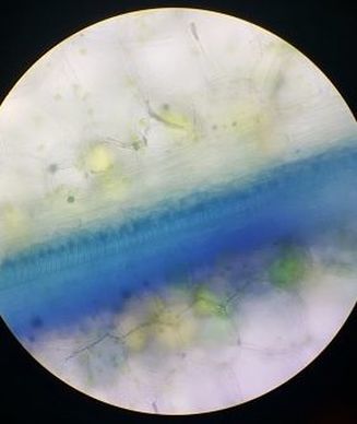 Figure 2: Longitudinal-section of snake plant (Sansevieria trifasciata) stained with TBO, Magnification: 40x, Shows elongated shape of fibers, lignified water-conducting cells; Photo credit: Melissa Clark; Slide Prep: Melissa Clark 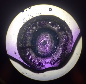 Figure 3: Cross-section of wax plant (Hoya carnosa) stained with CVA. Magnification: 40x, Shows epidermis, cortex, brachysclereids, the pith, and tracheids; Photo credit: Austin Wriggle; Slide Prep: Austin Wriggle Figure 3: Cross-section of wax plant (Hoya carnosa) stained with CVA. Magnification: 40x, Shows epidermis, cortex, brachysclereids, the pith, and tracheids; Photo credit: Austin Wriggle; Slide Prep: Austin Wriggle Tracheary Elements, Sclerids, and Parenchyma Tissue of Wax Plant (Hoya carnosa)
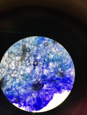
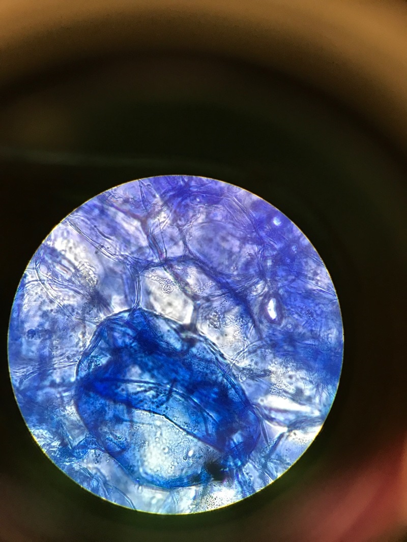
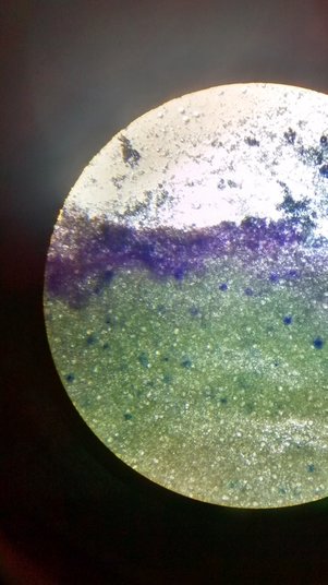
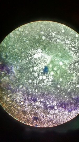
In order to examine the tracheary elements, sclerids, and parenchyma tissue in the wax plant, thin cross-sections of the stem were taken. These cross-sections were then stained with CVA. Observing the cross-section of the stem under a microscope, the CVA stained the sclerids and the water-conducting cells (tracheary elements) violet/blue . The outer ground tissue is made up of the parenchyma cells in the cortex and the ground tissue inside the ring of vascular tissue is called the pith. The parnechyma cells and the pith are differentiated into brachysclereids (Figure 3). Author: Austin Wriggle This past week in lab we got up close and personal with some familiar foods to learn about why they look and feel the way they do. The first plant we examined was the red cabbage. We were given cabbage leaves pretreated with digestive enzymes, which we then washed, filtered, and centrifuged. All of this was done in order to remove the cell wall leaving the protoplast behind. 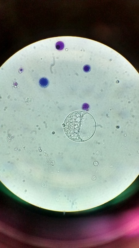 Red Cabbage (Brassica oleracea var. capitata f. rubra) protoplasts at 400x unstained. Photo and slide by Ally Kershner Red Cabbage (Brassica oleracea var. capitata f. rubra) protoplasts at 400x unstained. Photo and slide by Ally Kershner By removing the cell walls of the cabbage cells we were able to see them in a whole new light. While normally the cell wall would cause cells to be rigid and rectangular, these cells were circular. The outward pressure that is exerted by vacuoles is easy to see once the counterbalance of the cell wall is removed! Speaking of vacuoles, the vacuoles of the red cabbage are what gives it its distinctive color. As can be seen in the picture many of the smaller vacuoles are filled with blue and purple anthocyanin pigments. These pigments have been shown to have benefits for human health and may even help to prevent cancer. (On a personal note, over the summer I worked on a farm and was able to take home a lot of produce. One evening I made a stir fry with possibly the most potent and powerful purple cabbage in the world and immediately after had the first migraine I've had in years. I don't know that it was the anthocyanins but after that I definitely believe in the power of cabbages.) After we got a good look at the insides of a cell, and watched the cytoplasm move around for a while, we exposed our protoplasts to several different types of solution to see how they would react. Normally a plant cell, unlike an animal cell, has the cell wall to shield them so they can be a bit more resistant to things like changes in the concentration of solutes around them. However, because protoplasts don't have a cell wall, we thought that they would react more along the lines of animal cells. 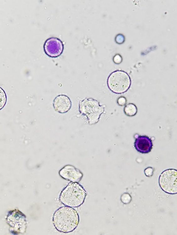 Red Cabbage (Brassica oleracea var. capitata f. rubra) protoplasts at 400x unstained with salt added. Slide and photo by Taylor Bates. Our predictions were right, and the protoplasts reacted visibly to the different solutions that we tried. Pictured about is how they shriveled up after being treated with salt, but we also exposed them to pure water (some of them exploded) and detergent (membranes were basically melted away, but not very quickly or anything). As well as looking at the insides of cabbages, last week we also covered the insides of other edible plants. Specifically we looked at the ground tissue of pears and avocados. We observed their sclerenchyma cells, which are what gives these fruits their texture. We stained both the pear and the avocado with TBO, which stained the lignified cell walls of the sclerenchyma blue so they were easy to see.
When we looked at the pear, it was easy to see that there were a lot of sclerenchyma cells as basically the whole sample was tinted blue. These are the brachysclereids or "stone cells" that you can feel when you eat pears. It was neat to be able to see the pit canals that went through the cell walls, especially when we looked at 400x magnification. It definitely explained why pears have the texture that they do!
We also stained avocado with TBO in order to look at its sclerenchyma cells. Like the pear, it had brachysclereids, but it had fewer of them and they were more sprinkled throughout the tissue. In the images above, the brachysclereids are the dark blue dots. Overall the avocado cells seemed a lot softer and blobbier looking, while the pear cells had more clearly defined borders. Avocados are much creamier and softer than pears in general so this makes sense. In fact I would rarely describe an avocado as gritty at all and previously wouldn't have compared it to a pear in any way.
All in all, it was very cool to get to look at some common fruits and veggies under the scope! I really like looking at things that I actually eat because it's knowledge that is directly connected to my life outside of class. Out of everything that we looked at last week, I think I was most interested in the avocado slide because it wasn't what I expected an avocado to look like. I definitely didn't think an avocado would be a great candidate for staining, or that avocado fruits had any particular structure besides just mush. I was happily surprised to be proved wrong! -Ally Kershner |
AuthorContent is created by students participating in the Plant Structure course at Oregon State University for Winter 2017. Archives
March 2017
Categories
All
|
 RSS Feed
