|
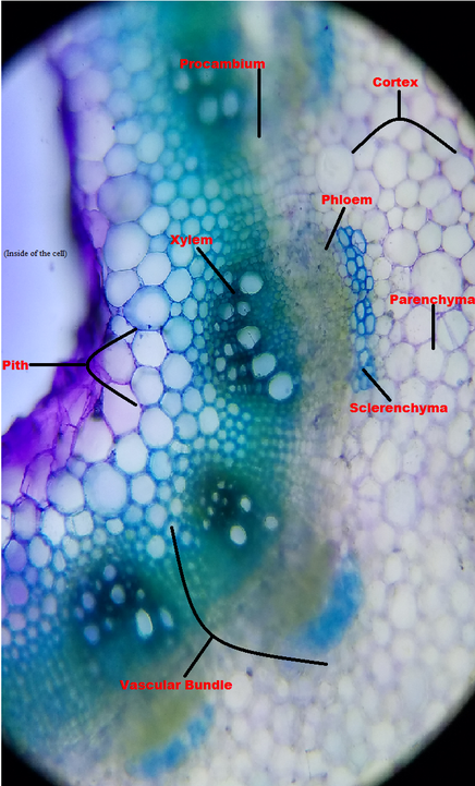
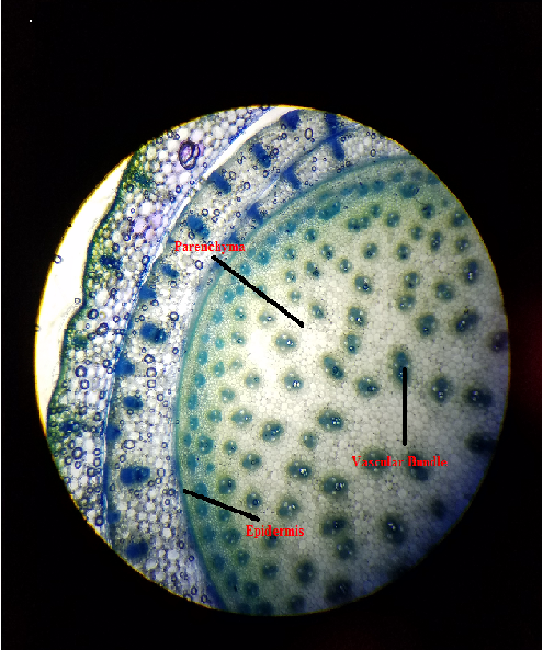
Oh man finally, I'm off of work. Now I can go to class and sit, I'm so tired of standing. Today's been a long day but now I have botany class! So here we are at lab and today's focus is about recognizing the tissues within stems and their functions, exploring the diversity of plant stems from different habitats, and seeing the difference between a monocot and a dicot plant. We did multiple cross sections of different types of plants. Let me tell you what! Cross sections are not easy. In order to get the best possible result you need to be able to cut the stem very thin. The problem for me is that my hand shakes too much, so it took me a couple tries to get a perfect cross section. One of my best cross sections is the broad bean stem, which can be seen in figure 1. This image was prepared and stained with Toluidine Blue O (TBO), and that's the reason we are able to see different colors and easily distinguish the different structures in this plant stem. The obvious thing you can notice between Figure 1 and Figure 2 is the complexity of a dicot structure. A distinct separation, that looks like a river that cuts through the forest, is called procambium. This separates the pith and the cortex of the stem. Not only that, comparing Figure 1 to Figure 2, their vascular bundle is very different from each other. In figure 1 you can see a separation between the xylem, which is responsible for transporting water and minerals, and the phloem, which is responsible for transporting food to the rest of the plant. But in figure 2 you can see that they're really close together, almost as if they were one. Now if we look closely at figure 1, you can see a blue stain on top of the phloem. That's what they call sclerenchyma, and we were told in class that this acts as a helmet and protects the phloem .
Now lets look at aquatic plants:
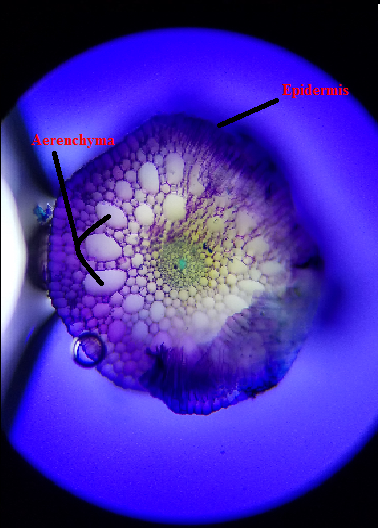
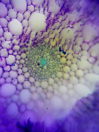
Figure 3: These are images of a waterweed (Elodea). This is a cross section of its stem, and was stained with TBO. This image was taken under a compound microscope at about 40x. The second image is a zoomed in version of the first image. (Prepared and photographed by Taylor) The plant structure in land plants compared to aquatic plants is very interesting. I've always thought that since they are all plants, their insides looks the same. I'm obviously wrong. There is a big difference. In aquatic plants I was able to learn that they contain these huge, easily seen air spaces throughout the stem called aerenchyma. Looking at figure 3 above, you can see what I am talking about. These air spaces are very important to aquatic plants because it provides buoyancy and it allows easier circulation of gases. Now after this lab I should be an expert at distinguishing the aquatic plants and terrestrial plants just by looking at their cross sections. Author: John P.
0 Comments
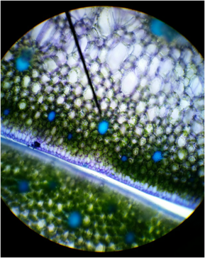 Figure 1: Cross-section of snake plant (Sansevieria trifasciata) stained with TBO, Magnification :100x, Shows sclerenchyma fibers, thickened secondary wall, parenchyma cells, and epidermis cells; Photo credit: Chris (Armstrong Plant Biology Lab) Slide prep: Chris (Armstrong Plant Biology Lab) Figure 1: Cross-section of snake plant (Sansevieria trifasciata) stained with TBO, Magnification :100x, Shows sclerenchyma fibers, thickened secondary wall, parenchyma cells, and epidermis cells; Photo credit: Chris (Armstrong Plant Biology Lab) Slide prep: Chris (Armstrong Plant Biology Lab) Introduction This past week in lab was all about learning and exploring the simple and complex tissues of plants. Our objectives were to recognize the three tissue systems of the plant body (ground, vascular, and dermal tissues), compare and contrast parenchyma, collenchyma, and sclerenchyma, identify water-conducting cells of the vascular tissue system and relate their structural features with their functions, and describe the characteristics of the epidermis, which we consider as a complex tissue. My primary focus for this lab was to prepare slides and observe the sclerenchyma fibers of a snake plant (Sansevieria trifasciata) by taking a cross-section and logitudinal-section of a leaf and observe the brachysclereids and tracheary elements of a wax pant (Hoya carnosa). Sclerenchyma Fibers of Snake Plant (Sansevieria trifasciata), Cross Section In order to examine the sclerenchyma fibers, a leaf was taken off of the snake plant. Using a razor blade, several thin cross-sections were taken from the leaf. A cross-section of the leaf was then stained in Toluidine Blue O (TBO) for about two minutes and then removed with a Kim wipe. Ethanol was added to the cross-section and then replaced with 20% CaCl and a cover slip. The reason the cross-section was stained with TBO was to observe the thickened secondary walls, which will be stained blue or blue-green in the presence of lignin. Vascular bundles, photosynthetic parenchyma cells of the mesophyll, and epidermis cells might have been also observed under the microscope (Figure 1). Once the cross-section was observed, a longitudinal-section was cut from the leaf and prepared on a slide stained in TBO. Observing the longitudinal-section of the snake plant leaf we were able to observe the elongated shape of the fibers, located in bundles. We were also able to see lignified water -conducting cells (Figure 2). 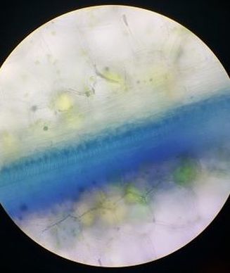 Figure 2: Longitudinal-section of snake plant (Sansevieria trifasciata) stained with TBO, Magnification: 40x, Shows elongated shape of fibers, lignified water-conducting cells; Photo credit: Melissa Clark; Slide Prep: Melissa Clark 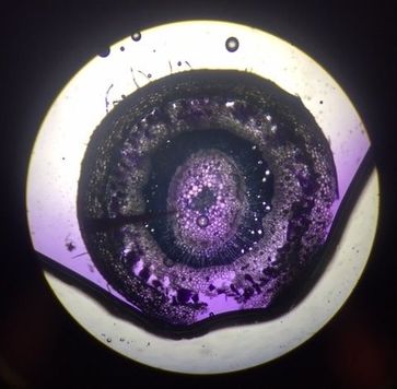 Figure 3: Cross-section of wax plant (Hoya carnosa) stained with CVA. Magnification: 40x, Shows epidermis, cortex, brachysclereids, the pith, and tracheids; Photo credit: Austin Wriggle; Slide Prep: Austin Wriggle Figure 3: Cross-section of wax plant (Hoya carnosa) stained with CVA. Magnification: 40x, Shows epidermis, cortex, brachysclereids, the pith, and tracheids; Photo credit: Austin Wriggle; Slide Prep: Austin Wriggle Tracheary Elements, Sclerids, and Parenchyma Tissue of Wax Plant (Hoya carnosa)
In order to examine the tracheary elements, sclerids, and parenchyma tissue in the wax plant, thin cross-sections of the stem were taken. These cross-sections were then stained with CVA. Observing the cross-section of the stem under a microscope, the CVA stained the sclerids and the water-conducting cells (tracheary elements) violet/blue . The outer ground tissue is made up of the parenchyma cells in the cortex and the ground tissue inside the ring of vascular tissue is called the pith. The parnechyma cells and the pith are differentiated into brachysclereids (Figure 3). Author: Austin Wriggle |
AuthorContent is created by students participating in the Plant Structure course at Oregon State University for Winter 2017. Archives
March 2017
Categories
All
|
 RSS Feed
