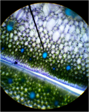
 Figure 1: Cross-section of snake plant (Sansevieria trifasciata) stained with TBO, Magnification :100x, Shows sclerenchyma fibers, thickened secondary wall, parenchyma cells, and epidermis cells; Photo credit: Chris (Armstrong Plant Biology Lab) Slide prep: Chris (Armstrong Plant Biology Lab) Figure 1: Cross-section of snake plant (Sansevieria trifasciata) stained with TBO, Magnification :100x, Shows sclerenchyma fibers, thickened secondary wall, parenchyma cells, and epidermis cells; Photo credit: Chris (Armstrong Plant Biology Lab) Slide prep: Chris (Armstrong Plant Biology Lab) Introduction This past week in lab was all about learning and exploring the simple and complex tissues of plants. Our objectives were to recognize the three tissue systems of the plant body (ground, vascular, and dermal tissues), compare and contrast parenchyma, collenchyma, and sclerenchyma, identify water-conducting cells of the vascular tissue system and relate their structural features with their functions, and describe the characteristics of the epidermis, which we consider as a complex tissue. My primary focus for this lab was to prepare slides and observe the sclerenchyma fibers of a snake plant (Sansevieria trifasciata) by taking a cross-section and logitudinal-section of a leaf and observe the brachysclereids and tracheary elements of a wax pant (Hoya carnosa). Sclerenchyma Fibers of Snake Plant (Sansevieria trifasciata), Cross Section In order to examine the sclerenchyma fibers, a leaf was taken off of the snake plant. Using a razor blade, several thin cross-sections were taken from the leaf. A cross-section of the leaf was then stained in Toluidine Blue O (TBO) for about two minutes and then removed with a Kim wipe. Ethanol was added to the cross-section and then replaced with 20% CaCl and a cover slip. The reason the cross-section was stained with TBO was to observe the thickened secondary walls, which will be stained blue or blue-green in the presence of lignin. Vascular bundles, photosynthetic parenchyma cells of the mesophyll, and epidermis cells might have been also observed under the microscope (Figure 1). Once the cross-section was observed, a longitudinal-section was cut from the leaf and prepared on a slide stained in TBO. Observing the longitudinal-section of the snake plant leaf we were able to observe the elongated shape of the fibers, located in bundles. We were also able to see lignified water -conducting cells (Figure 2). 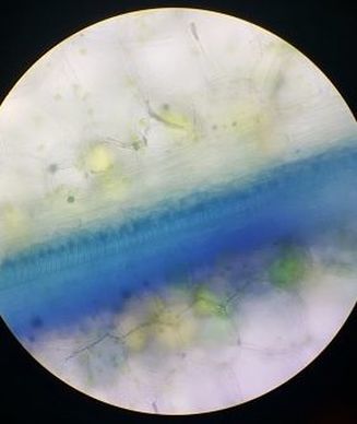 Figure 2: Longitudinal-section of snake plant (Sansevieria trifasciata) stained with TBO, Magnification: 40x, Shows elongated shape of fibers, lignified water-conducting cells; Photo credit: Melissa Clark; Slide Prep: Melissa Clark 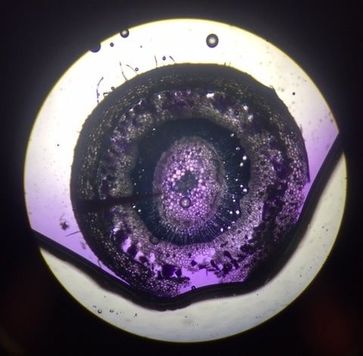 Figure 3: Cross-section of wax plant (Hoya carnosa) stained with CVA. Magnification: 40x, Shows epidermis, cortex, brachysclereids, the pith, and tracheids; Photo credit: Austin Wriggle; Slide Prep: Austin Wriggle Figure 3: Cross-section of wax plant (Hoya carnosa) stained with CVA. Magnification: 40x, Shows epidermis, cortex, brachysclereids, the pith, and tracheids; Photo credit: Austin Wriggle; Slide Prep: Austin Wriggle Tracheary Elements, Sclerids, and Parenchyma Tissue of Wax Plant (Hoya carnosa)
In order to examine the tracheary elements, sclerids, and parenchyma tissue in the wax plant, thin cross-sections of the stem were taken. These cross-sections were then stained with CVA. Observing the cross-section of the stem under a microscope, the CVA stained the sclerids and the water-conducting cells (tracheary elements) violet/blue . The outer ground tissue is made up of the parenchyma cells in the cortex and the ground tissue inside the ring of vascular tissue is called the pith. The parnechyma cells and the pith are differentiated into brachysclereids (Figure 3). Author: Austin Wriggle
0 Comments
Good evening! This week in Botany 313, we got to do some super interesting things. After talking things over with my lab partner and the other group doing a blog this week, I was given the responsibility of posting the epidermises of Tradescantia and a section of coleus (Plectranthus scutellarioides), so this post will focus mainly on those topics. In order to get a slide of the epidermises for Tradescantia, I started off with a leaf from the plant and with a little bending, snapping, and peeling I was able to successfully get a slide with both the upper and lower epidermises present. As shown in the images below, the stomata, guard cells, and pavement cells can clearly be seen on the lower epidermis. On the upper epidermis, only pavement cells can be seen as this type of plant does not have stomata on the upper epidermis. Stomata (plural of stoma) are openings in the outmost epidermal layer in plants that allow for the exchange of gases. They allow for a plant to retain water in times of drought or to increase rate of water loss in times when there is an excess of water. Guard cells allow for the opening or closing of the stomata with the aid of internal signaling (i.e. hormones), as well as external (i.e. The sun or rain). Pavement cells are simple cells with no real function other than protecting the cells below them. The purple pigmentation in these epidermal peels are from naturally occurring pigmentation within the plant. Taking a longitudinal section of coleus and finding the lignified tracheary elements was a bit more challenging. Simply getting sections that were thin enough requires some practice, and finding the tracheary elements, even more so. They are very easy to miss even with the stain! With a little patience, and maybe some help from Dr. L-P, I was able to find a few examples, as shown below. Tracheary elements, such as tracheids, aid in the conduction of water and minerals through the plant. 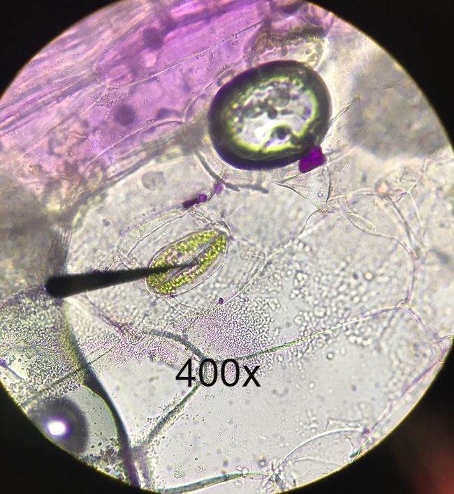 Lower epidermis of Tradescantia with no stains. Wet mount. Epidermal Peel. Arrow shown in image is from compound microscope. Arrow is pointing to stoma flanked by guard cells. The green dots in guard cells are chloroplasts. Cells surrounding guard cells are pavement cells. Photo credit: Michael Billard. Slide Prep credit: Michael Billard. 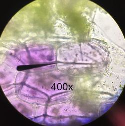 Upper epidermis of Tradescantia leaf with no stains. Wet mount. Epidermal Peel. Arrow shown is from compound microscope pointing to a pavement cell. Purple pigment shown is from naturally occurring pigments within the plant. No stomata can be seen on upper epidermis of plant leaf. Photo Credit: Michael Billard. Slide prep credit: Michael Billard. 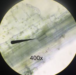 Longitudinal section of coleus (Plectranthus scutellarioides), stained with cresyl violet acetate (CVA) to show lignified tracheary elements. Wet mount. Arrow from compound microscope is pointing to lignified tracheary elements. Photo credit: Austin Wriggle. Slide prep credit: Michael Billard. Lesson Learned: Even with stains and a compound microscope, finding lignified tracheary elements can be very difficult. It can require cycling all the way through (and sometimes back) a focusing cycle, and even then, can be very easy to miss!
Author: Michael Billard |
AuthorContent is created by students participating in the Plant Structure course at Oregon State University for Winter 2017. Archives
March 2017
Categories
All
|
 RSS Feed
