|
Good evening! This week in Botany 313, we got to do some super interesting things. After talking things over with my lab partner and the other group doing a blog this week, I was given the responsibility of posting the epidermises of Tradescantia and a section of coleus (Plectranthus scutellarioides), so this post will focus mainly on those topics. In order to get a slide of the epidermises for Tradescantia, I started off with a leaf from the plant and with a little bending, snapping, and peeling I was able to successfully get a slide with both the upper and lower epidermises present. As shown in the images below, the stomata, guard cells, and pavement cells can clearly be seen on the lower epidermis. On the upper epidermis, only pavement cells can be seen as this type of plant does not have stomata on the upper epidermis. Stomata (plural of stoma) are openings in the outmost epidermal layer in plants that allow for the exchange of gases. They allow for a plant to retain water in times of drought or to increase rate of water loss in times when there is an excess of water. Guard cells allow for the opening or closing of the stomata with the aid of internal signaling (i.e. hormones), as well as external (i.e. The sun or rain). Pavement cells are simple cells with no real function other than protecting the cells below them. The purple pigmentation in these epidermal peels are from naturally occurring pigmentation within the plant. Taking a longitudinal section of coleus and finding the lignified tracheary elements was a bit more challenging. Simply getting sections that were thin enough requires some practice, and finding the tracheary elements, even more so. They are very easy to miss even with the stain! With a little patience, and maybe some help from Dr. L-P, I was able to find a few examples, as shown below. Tracheary elements, such as tracheids, aid in the conduction of water and minerals through the plant. 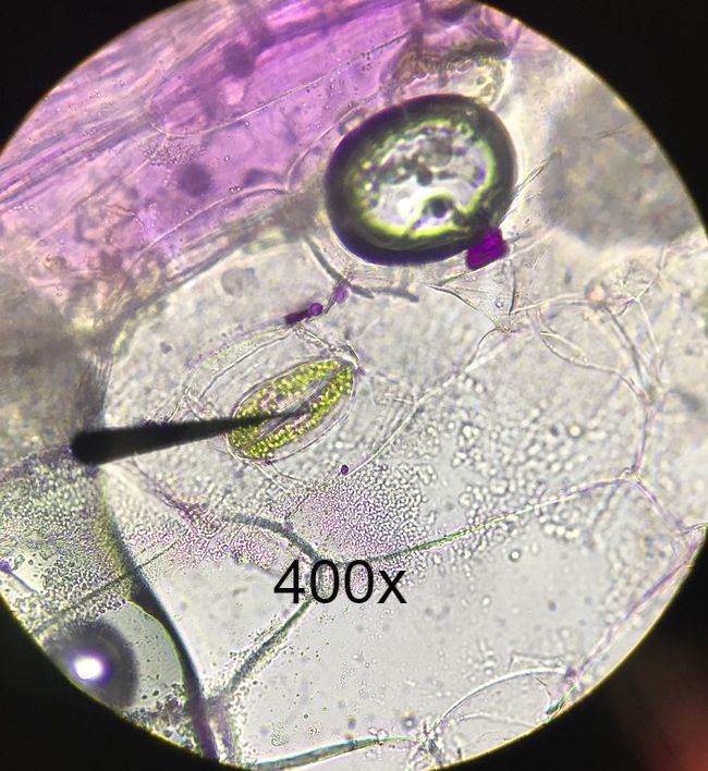 Lower epidermis of Tradescantia with no stains. Wet mount. Epidermal Peel. Arrow shown in image is from compound microscope. Arrow is pointing to stoma flanked by guard cells. The green dots in guard cells are chloroplasts. Cells surrounding guard cells are pavement cells. Photo credit: Michael Billard. Slide Prep credit: Michael Billard. 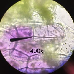 Upper epidermis of Tradescantia leaf with no stains. Wet mount. Epidermal Peel. Arrow shown is from compound microscope pointing to a pavement cell. Purple pigment shown is from naturally occurring pigments within the plant. No stomata can be seen on upper epidermis of plant leaf. Photo Credit: Michael Billard. Slide prep credit: Michael Billard. 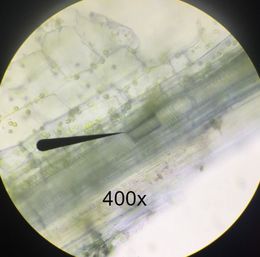 Longitudinal section of coleus (Plectranthus scutellarioides), stained with cresyl violet acetate (CVA) to show lignified tracheary elements. Wet mount. Arrow from compound microscope is pointing to lignified tracheary elements. Photo credit: Austin Wriggle. Slide prep credit: Michael Billard. Lesson Learned: Even with stains and a compound microscope, finding lignified tracheary elements can be very difficult. It can require cycling all the way through (and sometimes back) a focusing cycle, and even then, can be very easy to miss!
Author: Michael Billard
0 Comments
|
AuthorContent is created by students participating in the Plant Structure course at Oregon State University for Winter 2017. Archives
March 2017
Categories
All
|
 RSS Feed
