|
Last Tuesday in lab we studied about exploring the stomatal complexes of monocot leaf vs dicot leaf. We also learned about the internal structures of different leaves as well as their primary functions such as photosynthesis and transpiration. Stomatal Complexes of Monocot vs. Dicot Wheat cat grass is known Tritium vulgare, is monocot plant. Stomata of the wheat "cat-grass" (Tritium vulgare) consist of four cells, two guard cells and two subsidiary cells. The guard cells are specialized cells in the epidermis of leaves, stems and other organs that are used to control gas exchange. They are produced in pairs with a gap between them that form a stomatal pore. 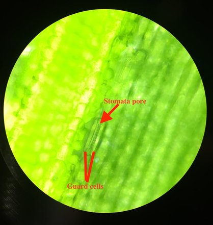 Figure 1: Epidermal peel of Wheat "cat-grass" (Tritium vulgar) unstained. Magnification: 400x. This picture shows two guard cells and stomata pore. Photo and Slide prep: Quyen Ta Figure 1: Epidermal peel of Wheat "cat-grass" (Tritium vulgar) unstained. Magnification: 400x. This picture shows two guard cells and stomata pore. Photo and Slide prep: Quyen Ta Broad bean is known Vicia faba, which is the dicot plant. Stomata plays a vital role in openings in the epidermal layer that allow for the exchange of gases. They allow for a plant to balance water inside and outside the cells. Guard cells allow for the opening or closing of the stomata with the internal hormone stimuli as well as external environmental factors. Pavement cells are simple cells with no real functions other than protecting the cells below them. Moreover, they help decrease water loss, and maintain an internal temperature. The most significant difference between the stomata of the monocots and the dicots is the shape of the guard cells. The monocot leaf has the narrow, dumbbell-shaped guard cells; whereas the dicot leaf has the pair-of-sausage shaped guard cells. Moreover, the monocot has the guard cells arranged in regular arrays, but the dicot has different paving. The monocot has stomata on both the upper and lower surface of the leaf. However, the dicot has stomata on the lower surface. 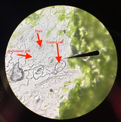 Figure 2: Epidermal peel of Broad Bean (Vicia faba) unstained. Magnification: 400x. Arrow is pointing to stomata by two guard cells. The green dots in guard cells are chloroplasts. Cells surrounding guard cells are pavement cells. Photographed by Max MacDonald and Slide prep by Quyen Ta Figure 2: Epidermal peel of Broad Bean (Vicia faba) unstained. Magnification: 400x. Arrow is pointing to stomata by two guard cells. The green dots in guard cells are chloroplasts. Cells surrounding guard cells are pavement cells. Photographed by Max MacDonald and Slide prep by Quyen Ta Cross-Section of Corn Leaf (Zea mays) Corn leaf (Zea mays) is monocot, has parallel veins. Moreover, spongy mesophyll is composed of parenchyma cells that contain chloroplast for photosynthesis. It also has air spaces for gas exchange and produces carbohydrates by photosynthesis. The upper and lower epidermis protect the leaf from water, sealing water inside and preventing parasite's attack. Xylem transports water into the leaf while phloem begins the sugar transport down to the roots. Veins is consisted of xylem and phloem, and a surrounding bundle sheath. 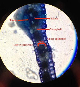 Figure 3: Cross-section of Corn Leaf (Zea mays) stained with TBO. Magnification: 400x. Photographed and slide pre by Quyen Ta Figure 3: Cross-section of Corn Leaf (Zea mays) stained with TBO. Magnification: 400x. Photographed and slide pre by Quyen Ta The internal structures of the monocot plants compared to the dicot plants made me surprised because I've always thought that their insides looks the same. However, there is a big difference. Guard cells of the monocot are narrow, dumbbell-shaped; but they are crazy-paving arrangement in the dicot. Stomata are located on both the upper and lower surface of the monocot leaf; whereas they are located only on the lower surface of the dicot leaf. During the lab, I felt difficulties in doing the cross-section of corn leaf because it needs a good skill technique to cut the cross-section. Finally, TA help me to finish the slide; the one I got that make me happy.
Submitted by Quyen Ta
0 Comments
This week we looked at a few leaves belonging to plants that have adapted to survive the niche in which they found themselves. Waterlilies have adapted to survive floating atop bodies of water, the rubber plant has adapted to survive tropical and equatorial climates, and the oleander has developed advantages for surviving in very dry climates.
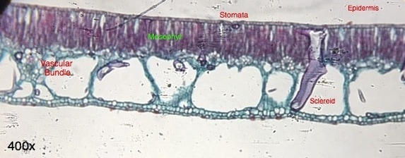
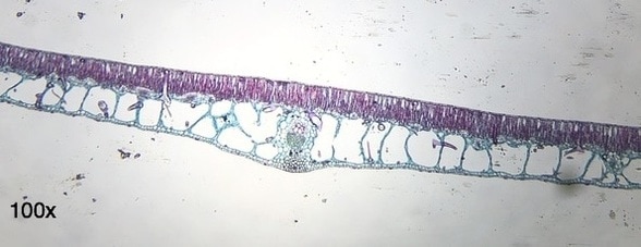
The Nymphaea (waterlily) leaf has stomata on top of their leaves instead of below to allow for increased air exchange and nutrient exchange; they do not have a defense against transpiration as other plants do in the form of guard cells. The loss due to transpiration is not a primary issue for the waterlily since it has extremely good access to water. The sclereid labeled above is meant as a support for the leaf; it helps tent up the leaf which allows for greater air exchange in the leaf air space and provides flotation for the pad. The cuticle of the leaf is quite thin and helps repel water from the stomata. -Chris Barrett 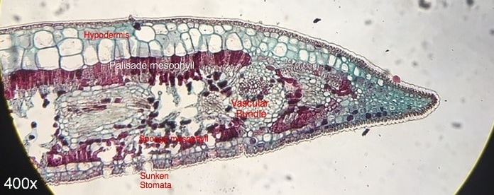 Ficus leaf, transverse section, Hematoxylin stain, 400x Magnification. Prepared by unknown and photographed by Chris Barrett 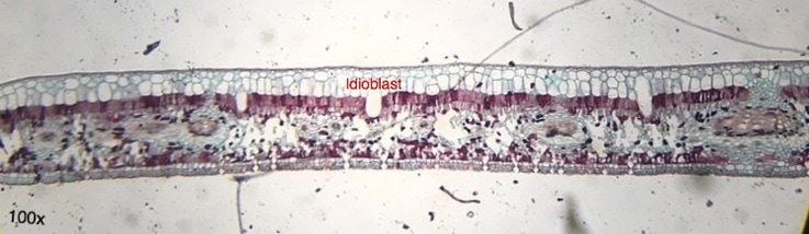 Ficus leaf, transverse section, Hematoxylin stain, 100x Magnification. Prepared by unknown and photographed by Chris Barrett The sunken stomata of the Ficus plant helps the plant retain water. By not being flush with the rest of the epidermis, the stomata allow water vapor to be released yet not be immediately blown away by wind, therefore retaining an amount of water for reabsorption. The hypodermis is quite thick on top of the palisade mesophyll cells, perhaps for protection from intense UV radiation. The sub-stomatal regions are quite large, allowing for greater gas exchange in the mesophyll cells. There are not any trichomes visible on this leaf section. This large open areas in the spongy mesophyll, the sunken stomata, and the thickened hypodermis point to this plant being able to survive very hot, and very sunny climates, perhaps in the equatorial region of the world. -Chris Barrett 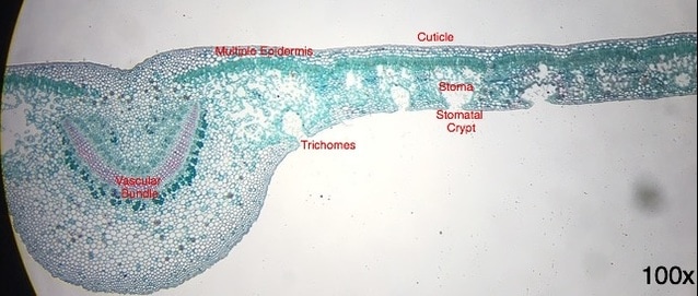 Nerium oleander leaf, transverse section, TBO stain, 100x Magnification. Prepared by unknown and photographed by Chris Barrett The adaptations of the Nerium oleander plant have allowed it to survive in very dry climates. These are evident through the presence of stomatal crypts, which contain multiple stoma positioned far away from outer line of the lower epidermis. There are also trichomes present near the openings of the stomatal crypts. Both the presence of the stomatal crypts and the trichomes located inside of them point to adaptations for survival in very dry climates. The increased presence of plant fibers in the leaves allows the leaf to maintain its shape even when its other cells are plasmolyzed in dry spouts. Its multiple epidermis and thick cuticle allows the plant to handle high ultra violet radiation.
Regardless of the fact that every part of the plant is toxic to humans and other animals, it is one of the most widely grown plants in the world due to its drought-resistance with uses including ornamental, medicine, and wind-blocking. The oleander plant is also the official flower of the city of Hiroshima as it was the first plant to flower there after the destruction of the city by nuclear blast. -Chris Barrett Good evening! This week in Botany 313, we got to do some super interesting things. After talking things over with my lab partner and the other group doing a blog this week, I was given the responsibility of posting the epidermises of Tradescantia and a section of coleus (Plectranthus scutellarioides), so this post will focus mainly on those topics. In order to get a slide of the epidermises for Tradescantia, I started off with a leaf from the plant and with a little bending, snapping, and peeling I was able to successfully get a slide with both the upper and lower epidermises present. As shown in the images below, the stomata, guard cells, and pavement cells can clearly be seen on the lower epidermis. On the upper epidermis, only pavement cells can be seen as this type of plant does not have stomata on the upper epidermis. Stomata (plural of stoma) are openings in the outmost epidermal layer in plants that allow for the exchange of gases. They allow for a plant to retain water in times of drought or to increase rate of water loss in times when there is an excess of water. Guard cells allow for the opening or closing of the stomata with the aid of internal signaling (i.e. hormones), as well as external (i.e. The sun or rain). Pavement cells are simple cells with no real function other than protecting the cells below them. The purple pigmentation in these epidermal peels are from naturally occurring pigmentation within the plant. Taking a longitudinal section of coleus and finding the lignified tracheary elements was a bit more challenging. Simply getting sections that were thin enough requires some practice, and finding the tracheary elements, even more so. They are very easy to miss even with the stain! With a little patience, and maybe some help from Dr. L-P, I was able to find a few examples, as shown below. Tracheary elements, such as tracheids, aid in the conduction of water and minerals through the plant. 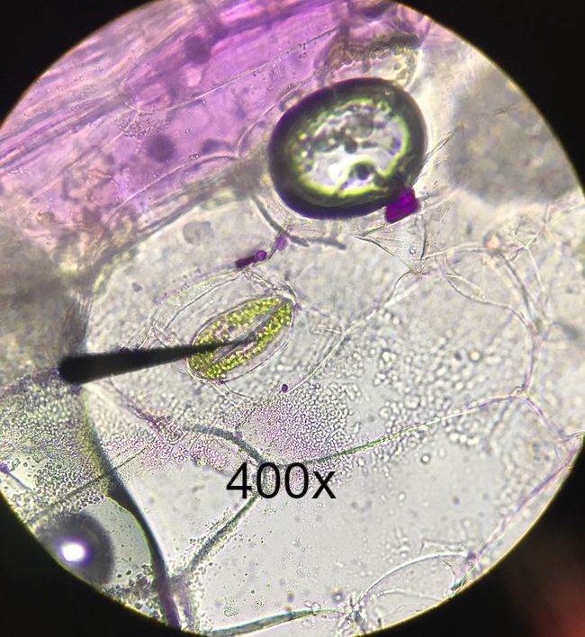 Lower epidermis of Tradescantia with no stains. Wet mount. Epidermal Peel. Arrow shown in image is from compound microscope. Arrow is pointing to stoma flanked by guard cells. The green dots in guard cells are chloroplasts. Cells surrounding guard cells are pavement cells. Photo credit: Michael Billard. Slide Prep credit: Michael Billard. 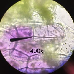 Upper epidermis of Tradescantia leaf with no stains. Wet mount. Epidermal Peel. Arrow shown is from compound microscope pointing to a pavement cell. Purple pigment shown is from naturally occurring pigments within the plant. No stomata can be seen on upper epidermis of plant leaf. Photo Credit: Michael Billard. Slide prep credit: Michael Billard. 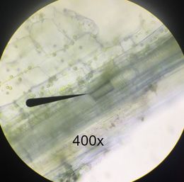 Longitudinal section of coleus (Plectranthus scutellarioides), stained with cresyl violet acetate (CVA) to show lignified tracheary elements. Wet mount. Arrow from compound microscope is pointing to lignified tracheary elements. Photo credit: Austin Wriggle. Slide prep credit: Michael Billard. Lesson Learned: Even with stains and a compound microscope, finding lignified tracheary elements can be very difficult. It can require cycling all the way through (and sometimes back) a focusing cycle, and even then, can be very easy to miss!
Author: Michael Billard |
AuthorContent is created by students participating in the Plant Structure course at Oregon State University for Winter 2017. Archives
March 2017
Categories
All
|
 RSS Feed
