Posted by Amber Eaton.
0 Comments
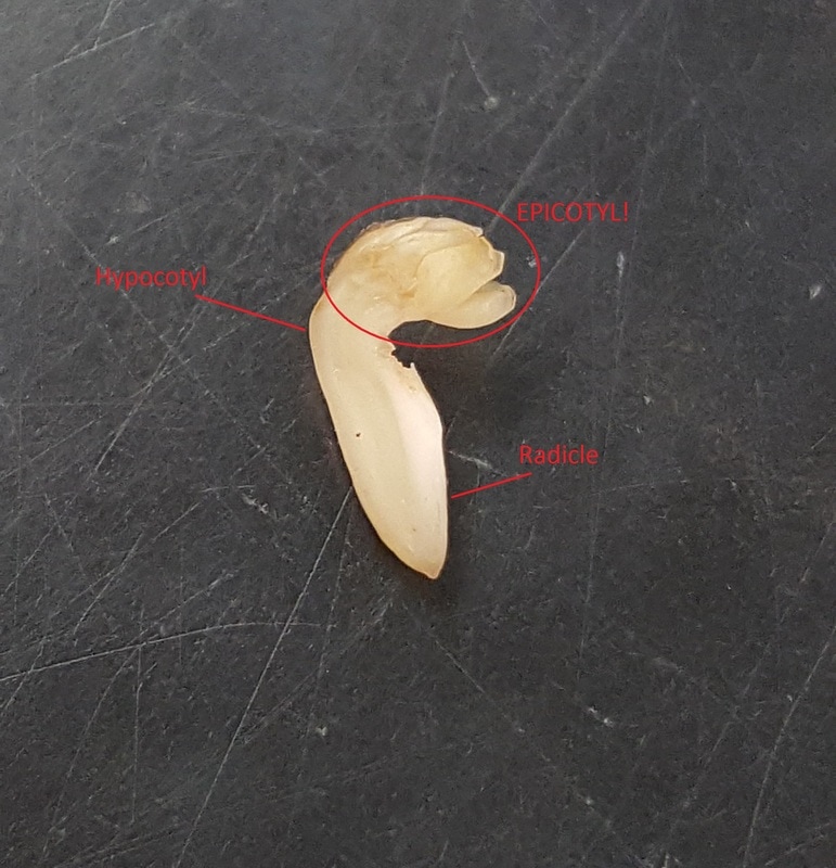 The embryo of Vicia fava, including the hypocotyl, the radicle, and the sneaky epicotyl. Prepared by Melissa Clark and Cierra Walker. Photo by Melissa Clark The embryo of Vicia fava, including the hypocotyl, the radicle, and the sneaky epicotyl. Prepared by Melissa Clark and Cierra Walker. Photo by Melissa Clark The fava beans of Botany 313 were proud to say they were perfectly normal, thank you very much. They were the last beans you’d expect to cause issues in a lab class, because they were large and easy to observe. Ok guys, I’m sorry. I couldn’t resist the chance to combine my two favorite things: botany and Harry Potter. Can you blame me? Probably. Be warned that you will see more references to the best book series of all time throughout this blog post. The first week in the Plant Structure lab was spent exploring seed anatomy, seed composition, seedling anatomy, and plant cell structures. My job is to tell you all about the fava bean, so let’s get to it. A seed is an incredible evolutionary development that, as Dr. L-.P. likes to say, “allows a plant to travel through time and space”. When a flower becomes pollinated, a plant begins to shuttle a great amount of nutrition to the now-developing plant embryo. As the embryo develops, so does the seed, providing protection in the form of a seed coat and a nutrient source, in the form of either endosperm or cotyledons, for the embryo after the seed has been removed from the mother plant. The embryo is now relatively safe to travel via a number of different dispersal methods that are specific to each plant species. Once they arrive in their new location, the seed has mechanisms that keep it in a quiescent, or inactive, state until external conditions are right for germination and seedling survival. So there it is; space-time travel without a Time Turner! The seed coat of our model fava bean was dark brown and surprisingly tough. The outside of the seed also has scars from fertilization and attachment to the mother plant, called the micropyle and the hilum respectively. When the seed coat was sliced off, the cotyledons were exposed. The energy source for the fava bean during germination is the cotyledons, not the endosperm as most are taught in general biology. Nestled between the cotyledons was the embryo, with its pre-stem tissue, called the hypocotyl, and its first root-like tissue, called the radicle, easily visible to the naked eye. Now, at this point in the lab, everyone is diligently searching for the various seed structures presented in the lab procedure. The structure that was eluding everyone was the epicotyl, a small, frilly region of the embryo on the hypocotyl. The epicotyl was so elusive, in fact, that Dr. L.-P. had to call off the search and tell the class that this structure probably wouldn’t be seen in our fava beans. So naturally, I channeled my inner Hermione and took this as a challenge. I closely scrutinized my bean halves and by holding the bean way too close to my face and focusing until my eyes almost crossed, I was able to see it! I had found the epicotyl! It was disappointing to see that the next step of the lab procedure was to dissect the embryo even further, meaning I would have to destroy this tiny structure, but you know how the saying goes: For science! So I chopped up the poor little embryo and soaked it in chemical stains in order to observe its composition and now dying metabolism. Four slices of both the cotyledon and the embryo were made. With one set of slices as a control, the stains were applied to the cut surface and allowed to sit for a few minutes to fully dye. The stains used were Lugol’s iodine for presence of starch, Sudan IV for presence of oils, and methylene blue for active respiration. As the photo shows, both the embryo and the cotyledon contained starch, but the cotyledon was stained darker than Voldemort’s heart, which is to say it had a higher concentration of starch. Again, both slices were stained by the Sudan IV, but more apparent staining happened to the cotyledon. Both of these results are to be expected, as the cotyledon stores starches and oils as nutrition for early plant development. The next stain is a little harder to distinguish in the picture because there was a little excess methylene blue left on the cotyledon (oops). The embryo is definitely stained a deeper blue, indicating that active respiration is occurring, although not for much longer. Poor baby. 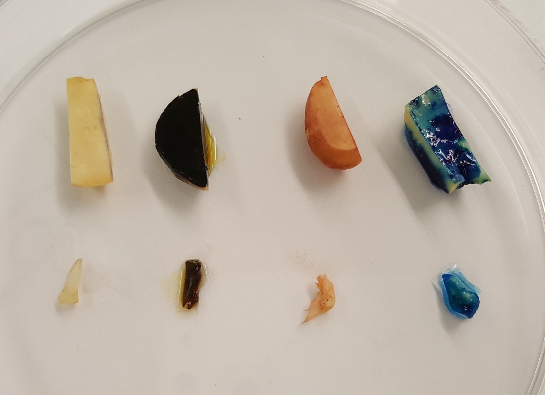 Staining of V. fava from left to right: control (no stain), Lugol's iodine (I2KI, darkness), Sudan IV, and methylene blue. Prepared by Melissa Clark and Cierra Walker. Photo by Melissa Clark The next model (like supermodel; this plant was beautiful), was a three-week-old fava bean seedling. A seed had germinated in a container of water and grown into a seedling with every piece of its morphology exposed. There was no soil to hide the seed or the roots and shoot protruding from it. “Naked” plants are very aesthetically pleasing. A seedling can have some remnants of embryonic structures; usually the cotyledons and the hypocotyl. These structures aren’t visible on fava beans, though, because they undergo hypogeal germination, or germination that occurs beneath the soil and within the seed coat. The alternative to this is epigeal germination, during which a seed germinates and the cotyledons expand out of the seed coat and above ground to begin photosynthesis. In the case of the fava bean, photosynthesis occurs in the first true leaves. I tried my hardest to create beautiful, botanically accurate diagrams for you guys. It didn’t work out so well, though. Everything is labeled and you can probably get the vague sense that these could be drawings of plants. Please have as much mercy on this picture as the TA who graded it. 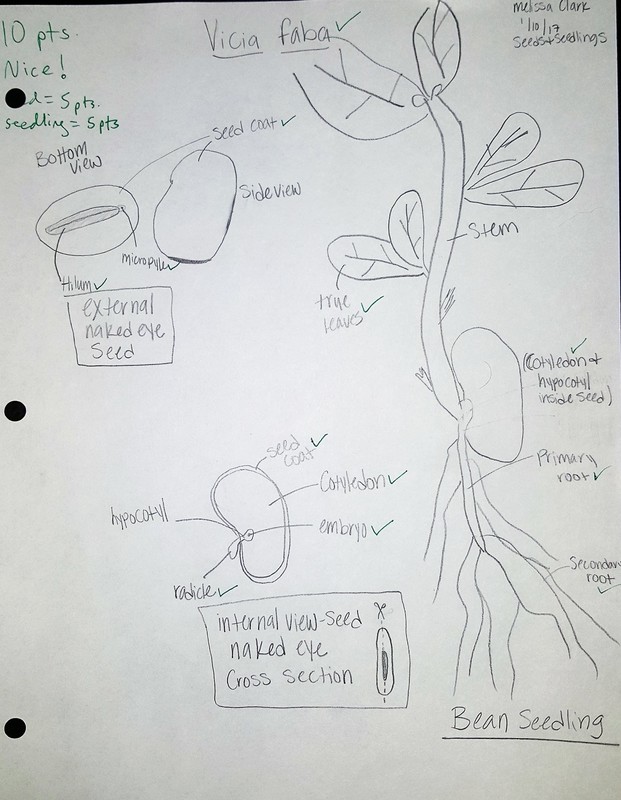 Beautifully drawn and accurately labeled fava bean and seedling, by Melissa Clark Now, as a gift for enduring that last picture, here’s a fava bean taking on the form of an angry bird.  Bean? Bird? Either way, it's angry. Photo by Melissa Clark Author: Melissa Clark |
AuthorContent is created by students participating in the Plant Structure course at Oregon State University for Winter 2017. Archives
March 2017
Categories
All
|
 RSS Feed
