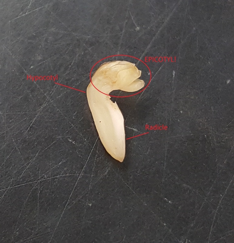
 The embryo of Vicia fava, including the hypocotyl, the radicle, and the sneaky epicotyl. Prepared by Melissa Clark and Cierra Walker. Photo by Melissa Clark The embryo of Vicia fava, including the hypocotyl, the radicle, and the sneaky epicotyl. Prepared by Melissa Clark and Cierra Walker. Photo by Melissa Clark The fava beans of Botany 313 were proud to say they were perfectly normal, thank you very much. They were the last beans you’d expect to cause issues in a lab class, because they were large and easy to observe. Ok guys, I’m sorry. I couldn’t resist the chance to combine my two favorite things: botany and Harry Potter. Can you blame me? Probably. Be warned that you will see more references to the best book series of all time throughout this blog post. The first week in the Plant Structure lab was spent exploring seed anatomy, seed composition, seedling anatomy, and plant cell structures. My job is to tell you all about the fava bean, so let’s get to it. A seed is an incredible evolutionary development that, as Dr. L-.P. likes to say, “allows a plant to travel through time and space”. When a flower becomes pollinated, a plant begins to shuttle a great amount of nutrition to the now-developing plant embryo. As the embryo develops, so does the seed, providing protection in the form of a seed coat and a nutrient source, in the form of either endosperm or cotyledons, for the embryo after the seed has been removed from the mother plant. The embryo is now relatively safe to travel via a number of different dispersal methods that are specific to each plant species. Once they arrive in their new location, the seed has mechanisms that keep it in a quiescent, or inactive, state until external conditions are right for germination and seedling survival. So there it is; space-time travel without a Time Turner! The seed coat of our model fava bean was dark brown and surprisingly tough. The outside of the seed also has scars from fertilization and attachment to the mother plant, called the micropyle and the hilum respectively. When the seed coat was sliced off, the cotyledons were exposed. The energy source for the fava bean during germination is the cotyledons, not the endosperm as most are taught in general biology. Nestled between the cotyledons was the embryo, with its pre-stem tissue, called the hypocotyl, and its first root-like tissue, called the radicle, easily visible to the naked eye. Now, at this point in the lab, everyone is diligently searching for the various seed structures presented in the lab procedure. The structure that was eluding everyone was the epicotyl, a small, frilly region of the embryo on the hypocotyl. The epicotyl was so elusive, in fact, that Dr. L.-P. had to call off the search and tell the class that this structure probably wouldn’t be seen in our fava beans. So naturally, I channeled my inner Hermione and took this as a challenge. I closely scrutinized my bean halves and by holding the bean way too close to my face and focusing until my eyes almost crossed, I was able to see it! I had found the epicotyl! It was disappointing to see that the next step of the lab procedure was to dissect the embryo even further, meaning I would have to destroy this tiny structure, but you know how the saying goes: For science! So I chopped up the poor little embryo and soaked it in chemical stains in order to observe its composition and now dying metabolism. Four slices of both the cotyledon and the embryo were made. With one set of slices as a control, the stains were applied to the cut surface and allowed to sit for a few minutes to fully dye. The stains used were Lugol’s iodine for presence of starch, Sudan IV for presence of oils, and methylene blue for active respiration. As the photo shows, both the embryo and the cotyledon contained starch, but the cotyledon was stained darker than Voldemort’s heart, which is to say it had a higher concentration of starch. Again, both slices were stained by the Sudan IV, but more apparent staining happened to the cotyledon. Both of these results are to be expected, as the cotyledon stores starches and oils as nutrition for early plant development. The next stain is a little harder to distinguish in the picture because there was a little excess methylene blue left on the cotyledon (oops). The embryo is definitely stained a deeper blue, indicating that active respiration is occurring, although not for much longer. Poor baby. 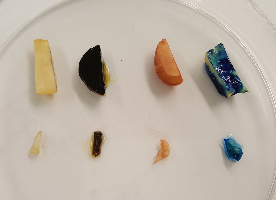 Staining of V. fava from left to right: control (no stain), Lugol's iodine (I2KI, darkness), Sudan IV, and methylene blue. Prepared by Melissa Clark and Cierra Walker. Photo by Melissa Clark The next model (like supermodel; this plant was beautiful), was a three-week-old fava bean seedling. A seed had germinated in a container of water and grown into a seedling with every piece of its morphology exposed. There was no soil to hide the seed or the roots and shoot protruding from it. “Naked” plants are very aesthetically pleasing. A seedling can have some remnants of embryonic structures; usually the cotyledons and the hypocotyl. These structures aren’t visible on fava beans, though, because they undergo hypogeal germination, or germination that occurs beneath the soil and within the seed coat. The alternative to this is epigeal germination, during which a seed germinates and the cotyledons expand out of the seed coat and above ground to begin photosynthesis. In the case of the fava bean, photosynthesis occurs in the first true leaves. I tried my hardest to create beautiful, botanically accurate diagrams for you guys. It didn’t work out so well, though. Everything is labeled and you can probably get the vague sense that these could be drawings of plants. Please have as much mercy on this picture as the TA who graded it. 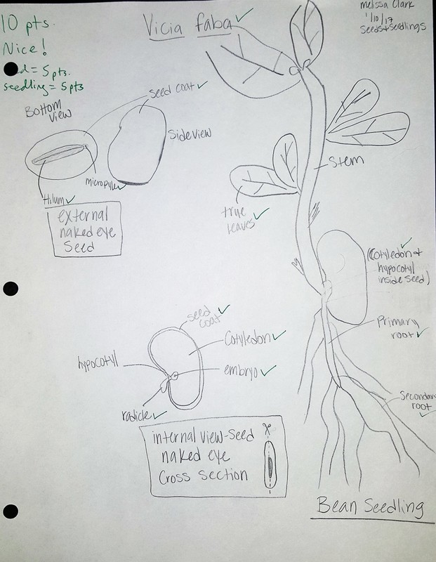 Beautifully drawn and accurately labeled fava bean and seedling, by Melissa Clark Now, as a gift for enduring that last picture, here’s a fava bean taking on the form of an angry bird. 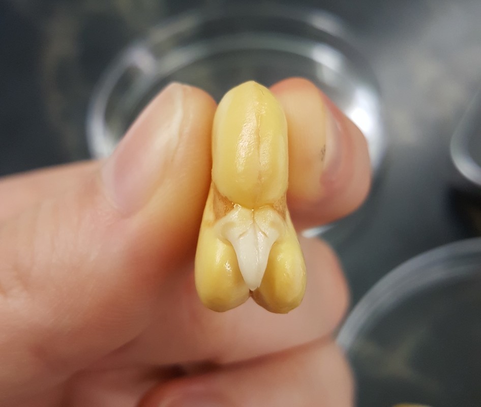 Bean? Bird? Either way, it's angry. Photo by Melissa Clark Author: Melissa ClarkAmateur Botanist Explores Microscopic Magic Crystals, Extra-Hairy Plants, and Alien Spores1/15/2017
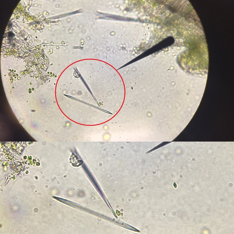
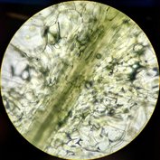
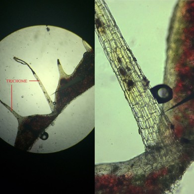 Begonia Trichomes. Left: epidermal hairs at 40x magnifcation. Right: Base of epidermal hair at 400x magnification, unstained, longitudinal section. Photo taken by Amy K. Begonia Trichomes. Left: epidermal hairs at 40x magnifcation. Right: Base of epidermal hair at 400x magnification, unstained, longitudinal section. Photo taken by Amy K. I am a house plant addict. As in, turn-my-apartment-into-a-jungle-and-call-it-home kind of addict. I could use the phrase ‘house plant enthusiast’, but it doesn’t quite capture the depth of my affection for these organisms. So imagine my excitement upon seeing a table full of some of my favorites placed at the front of the lab room. Imagine again my slight adrenaline rush in in learning that we were going to study them under microscopes - exploring their cells, traveling to far off places unreachable by the naked eye. I was stoked. For starters, I had a quick *heart eyes* moment with one of the specimens, a majestic (and I do mean majestic) Begonia before diving in to the task at hand: trichome collection and examination. Trichomes are hair-like structures that cover the leaves and stem of the plant; they come in many shapes and sizes and serve a variety of functions. In some plants, trichomes are coated in sticky substances capable of trapping insects, preparing them for chemical digestion. In others, the hairs prevent herbivory by means of painful chemical injection. Begonias are more ‘sunshine and daisies’ when it comes to their epidermal hairs, most likely serving to collect moisture from the air or prevent herbivory by having an unpleasing texture. After producing a wet mount slide, I was surprised to see a difference in the cellular shape of the trichome relative to the those of the leaf it was attached to. Where leaf cells were circular in shape, cells of the trichomes were rectangular. Furthermore, fuzzy deep red pigments were mixed with slight pockets of green – an act of Christmas regurgitating its holiday sprinkles all over this plant or anthocyanins masking chlorophyll pigments? I’ll let you decide. Next up: examining raphide crystals found within Tradescantia zebrina (aka “Inchplant”) and druse crystals formed in an unknown species of Begonia via wet mount slides. These structures are products of excess inorganic particles (most often calcium salts) being deposited into the vacuole in crystalline form. The raphide crystals were easy enough to find, often found in clusters mixed with cell matter. However, searching for druse crystals felt like searching for Waldo in a sea of cartoon people; you find him once, painstakingly, and then you can’t find him again.
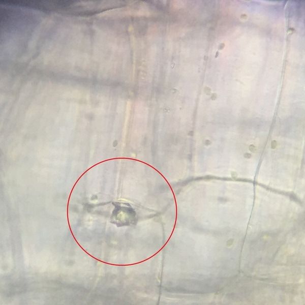
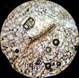
My favorite part of this lab reminded me of one of my all-time favorite films, The Fifth Element, which revolves around the “untraditional hero saves planet” trope. Bruce Willis plays Korben Dallas, an ex-galactic special forces operative turned cab driver of the 23rd century, who attempts to save the world from a giant ball of talking fire in space. I can't make this stuff up, guys - its cinematic gold. That giant ball of talking fire, who calls himself "Mr. Shadow", looked pretty similar to the Ceratopteris richardii spore under my microscope (in my humble opinion). Under 400x magnification, the surface ridges on this tiny sphere give it the look of some far off planet in outer space. In reality, it comes from an aquatic fern found on Planet Earth, dubbed the “C-Fern”. Our mission: examine the spores under a microscope and sow into prepared culture plates containing agar and mineral nutrients, where it will be examined further in the coming weeks. 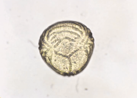 Epidermal ridges on C-Fern - 400x magnification, unstained, wet mount. GIF made by Amy K.  Mr. Shadow aka Giant Talking Ball of Fire. GIF made by Amy K. Author: Amy KHouse plant addict. Believer in Himalayan Salt lamps. Enjoys the little things in life like popcorn and vegan marshmallows. Submitted by Brandon Quann
Hello and welcome to my very first blog post ever. I’ve decided to add my own twist to my post so I've included some of my favorite Kanye West instrumentals to add to the "ambiance" of my post, to start the playlist press play down below.
This week in lab we learned about the inner workings of seeds, seedlings, and plant cells. We carried out experiments by conducting dissections, staining plant tissues with various biological stains, preparing and viewing microscope slides, and illustrating our findings. In our lab we observed numerous plants and tissues types, however I will be focusing on our experiments with Elodea cells as well as plastids within bell peppers.
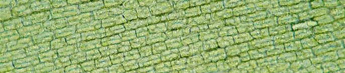
Elodea cells under 100x magnification. Photo by Taylor Bates
Who knew pondweed could be so interesting?
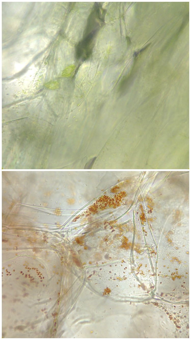
It has been quite some time since my high school biology class so when I saw that we would be studying Elodea cells in lab this week I had a trip down memory lane to my first encounter with the plant. Our observations began with making a simple wet mount of the leaf and viewing it under 100x and 400x magnification. Once we had our slides made we began searching for and sketching trichomes, chloroplasts, nuclei, vacuoles, cell walls, and any other organelles we could find.
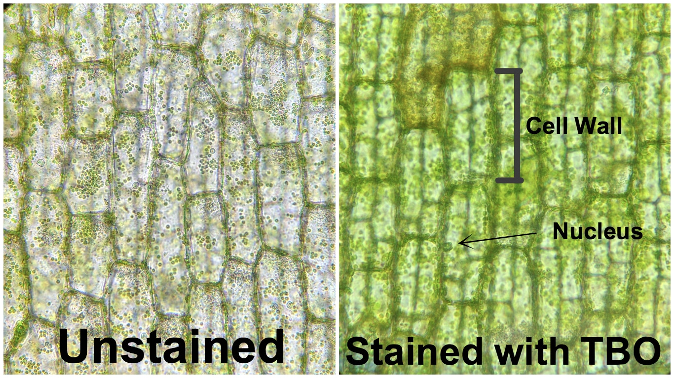
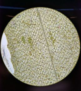
Elodea leaves were then stained with a different biological stain called Toluidine Blue O (TBO). TBO creates a variety of colors when in contact with different tissue types, the structures/ organelles and their corresponding stain color are in the following list: pectin found in cell walls, cell wall tissue, and nuclei. Based on these corresponding colors it is apparent that most abundant tissue found in Elodea leaves is from the cell wall (see photo above). A few nuclei are also visible from the stain however this is the first time I have used TBO so I am not sure if I allowed the stain enough time to work to its full potential.
How much do Plastids cost?
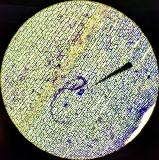
dependent on environmental conditions or maturity of the fruit. These 3 different types of plastids have their own individual phenotypic differences as well as different functional roles in the cell. The photo above on the left shows the tissue of a green bell pepper with high levels of chloroplasts, these chloroplasts are the main site of photosynthesis and appear green because of the chlorophyl inside. However if the fruit is left on the plant to mature for a longer period of time the chloroplasts will be converted into chromoplasts. These chromoplasts are non-photosynthetic and work to synthesize and store pigments called carotenes, which are yellow, orange, or red in color (depending on how long they are left to mature). The conversion of chloroplasts into chromoplasts can be seen in the photo above on the right which shows a tissue sample of a red bell pepper full of red chromoplasts.
|
AuthorContent is created by students participating in the Plant Structure course at Oregon State University for Winter 2017. Archives
March 2017
Categories
All
|
 RSS Feed
